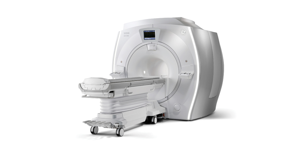
More Accurate Test for Detection of Prostate Cancer Now Available in MRI
According to the American Cancer Society, prostate cancer is the 2nd most common cancer in American men behind skin cancer. Prostate cancer is the 2nd leading cause of cancer death in men behind lung cancer. One man in 6 will be diagnosed with prostate cancer during his lifetime.
Given these statistics, attempts to develop an accurate and reliable method to both screen for and diagnose prostate cancer have met with limited success. The PSA blood test is the best indicator for the presence of prostate cancer. Unfortunately, the PSA can be elevated in other conditions including prostate inflammation. The current method of Trans Rectal Ultrasound (TRUS) biopsy of the prostate to diagnose cancer in men with an elevated PSA is both inaccurate and misleading leaving the utility of the biopsy in question. TRUS biopsies in patients with an elevated PSA find insignificant cancer 40% of the time, miss significant cancer entirely 40% of the time and in those patients in which it finds cancer, it misses the most aggressive portion of the cancer 40% of the time. These results are behind the recommendation of many professional organizations to abandon the use of PSA as a screening test for prostate cancer.
MRI Offers Prostate Cancer Detection Rates Around 90%
However, MRI of the prostate has now emerged as a new test to both accurately diagnose and localize prostate cancer. MRI has been used successfully to diagnose neurological and musculoskeletal conditions for many years. Now, MRI is being used to generate detailed images of the prostate through the acquisition of high resolution images combined with dynamic contrast enhanced images to make the accurate diagnosis of prostate cancer. Multi-parametric high field 3T MRI has been shown to detect prostate cancer with 90% accuracy. As important, if the MRI exam is normal, there is a less than 3% chance that significant disease is present. MRI has emerged as a successful adjunct to TRUS biopsy diagnosis of prostate cancer by providing accurate information on the location and severity of disease, thereby aiding urologists to more precisely target their biopsies.
Because 3T MRI images the entire prostate and surrounding structures, it can detect high grade and multifocal cancers and thereby determine the full extent of disease. It is also the most accurate method of diagnosing and following low grade tumors which don’t require biopsy or aggressive treatment.
NorCal Imaging of Walnut Creek is using the most advanced Multi-parametric high field 3T MRI technology in the detection of prostate cancer. The scan takes less than one hour and there is no rectal coil. This imaging process allows for the most accurate detection, characterization, localization and staging of prostate cancer for TRUS guided biopsies, treatment and follow up.
NorCal Imaging is one of the only outpatient imaging providers in the Bay Area currently offering 3T prostate MRI with multi-parametric Invivo computer assisted diagnosis. This technology is also essential in making the decision for active surveillance of low grade prostate tumors, thereby avoiding unnecessary biopsies.
Dr. Charles Fiske is a radiologist and Medical Director of NorCal Imaging in Walnut Creek. He specializes in oncologic imaging, diagnosis, staging and image guided interventions. For more information on the use of MRI for prostate cancer diagnosis and staging, call 925-937-6100