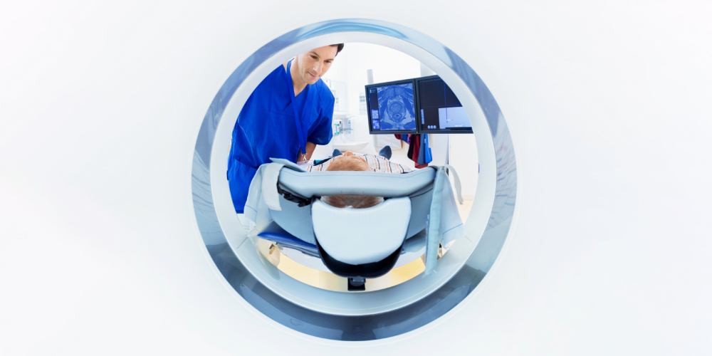
Multiparametric MRI is one of the latest and most effective tools employed to detect prostate cancer. It supplements ordinary MRI scans with additional imaging in order to provide radiologists with detailed anatomical information about the patient’s prostate gland. If a tumor is detected, multiparametric MRI allows radiologists to measure its size, map its position, determine its stage, estimate its Gleason score, and check whether it has spread beyond the prostate.
Multiparametric MRI Imaging Sequence
A multiparametric imaging sequence is comprised of three separate imaging techniques: T2-weighted MRI, diffusion-weighted MRI, and dynamic contrast-enhanced MRI.
T2-Weighted MRI
T2-weighted MRI has been described as the “workhorse” of multiparametric MRI.2 A standard MRI creates 3D images by manipulating water molecules in human tissue, using magnetic fields and radio pulses. By altering the length of the pulse, radiologists can highlight certain types of tissue in the human body. A T2 pulse is especially beneficial at imaging cancerous tissue because of differences in water content.
T2-weighted MRI lets doctors examine all the anatomical zones within the prostate: the peripheral zone (closest to the rectum), the transitional zone (around the urethra), and the central zone (around the ejaculatory ducts). Tumors in the peripheral zone are easiest to spot – they show up as dark spots against a bright background – while cancers in the transitional zone are more difficult to see. On T2-weighted images, they are like dark, smudged charcoal.
Most importantly, T2-weighted MRI provides enough detail for radiologists to examine the shape and size of the tumor, and determine its stage. It also allows physicians to see if the cancer has spread to the surrounding structures, such as the bladder, urinary sphincter, and seminal vesicles.
Diffusion-Weighted MRI
Diffusion-weighted MRI measures the motion of water molecules inside the body. The medical term is ADC (apparent diffusion coefficient). Cancers have a low ADC because they restrict the movement of water. Once a cancer has been identified, its ADC can help determine how aggressive it is. Low ADC is correlated with higher Gleason scores and greater patient risk.
Dynamic Contrast-Enhanced MRI
Dynamic contrast-enhanced MRI uses contrast agents, injected into the blood stream, to make bodily tissues more visible on a scan. Contrast agents are not radioactive. They affect the spin of water molecules inside the prostate, which is one of the main sources of contrast in MRI images. After the exam, the agent is eliminated naturally through the kidneys.
Dynamic contrast-enhanced MRI allows radiologists to measure blood flow through the prostrate. As tumors grow, they develop abnormal blood vessels that can be identified by tracking blood circulation. Cancers also absorb contrast agents more quickly than healthy tissue, making them stand out more on an MRI.
Because abnormal blood flow is also a characteristic of benign prostate conditions such as prostatitis and BPH nodules, dynamic contrast-enhanced MRI is a secondary imaging technique. Radiologists rely on it to increase their confidence in identifying lesions, to help finalize the Gleason score for indeterminate tumors, and to provide additional information when a patient’s primary scans are blurred or distorted. One area where dynamic contrast is especially useful: detecting small cancers invisible on T2 or diffusion MRIs.
Multiparametric MRI Facilities
Patients interested in multiparametric MRI should be aware of what type of equipment is being used to capture their images. Not all MRI machines are equally effective. Currently, the best in the field is the 3T MRI. However, many facilities still use the 1.5T MRI, which is half as strong and has a weaker signal-to-noise ratio.
3T MRI is the standard equipment used in RadNet facilities, although 1.5 Tesla MRIs may be used in some situations. Led by Dr. Princenthal, one of the chief experts in the field, our prostate cancer screening program is expanding access to multiparametric MRI to men across the nation.
References
- Robert Princenthal, MD. Adding Multiparametric MRI to Prostate Cancer Screening Will Save Lives and Money
- Ghai, S. and Haider, M. A. Multiparametric-MRI in Diagnosis of Prostate Cancer. Indian Journal of Urology. 2015 Jul-Sep; 31(3): 194–201.