Magnetic resonance imaging (MRI) uses powerful magnets to detect radiofrequency signals as they travel through the body. Using advanced computer software, these radiofrequency signals are translated into detailed digital visualizations of internal body parts, like the prostate gland.
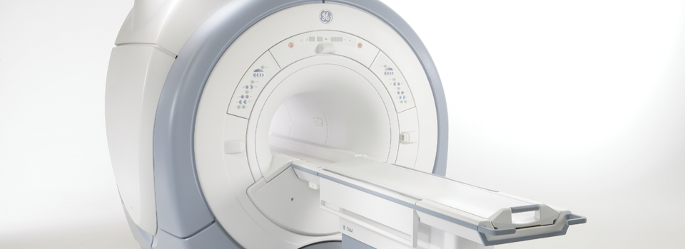
The field strength of magnets used in MRI machines are measured in ‘Teslas’ (T). More Teslas indicates a higher field strength. The field strength of magnets available to most patients has progressed over time from .2T to .7T to 1.2T to 1.5T. Today, 3T MRI is becoming more widely available and represents some of the most advanced imaging technology deployed in clinical settings.
3T MRI vs. 1.5T MRI in Prostate MRI
Although lower field strength MRIs are serviceable and appropriate for many imaging exams, 3T MRI produces higher-resolution images. Both 1.5T and 3T MRI are increasingly used with success in prostate imaging. However, 3T MRI prostate imaging appears to offer significant improvements in spatial resolution and local staging accuracy compared with 1.5T MRI. Benefits of 3T MRI include:
- Increased signal-to-noise ratio, which results in images that appear less grainy than those produced by lower field strength MRIs. This provides more accurate functional data during certain sequences.
- Increased spatial resolution, which results in images that are sharper and appear less pixelated than those produced by lower field strength MRIs. This offers more detailed anatomical images of the prostate gland.
- Increased temporal resolution, which results in shorter scan times than are possible with lower field strength MRIs. This can reduce the need for an endorectal coil, a device inserted into the rectum during prostate MRI to produce more detailed images.
To ensure patients receive best possible benefit from prostate MRI, RadNet only offers prostate MRI services at facilities equipped with 3T MRI.